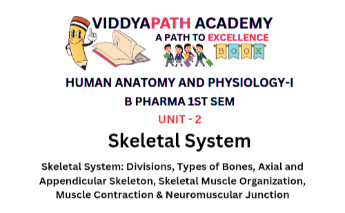
Skeletal System: Divisions, Types of Bones, Axial & Appendicular Skeleton, Skeletal Muscle Organization, Muscle Contraction & Neuromuscular Junction
Introduction: Teaching the Skeletal System the Right Way
The skeletal system is not just a rigid support structure. It is a dynamic, metabolically active system that protects vital organs, enables movement, produces blood cells, stores minerals, and works hand-in-hand with muscles and nerves.
In this in-depth, exam-oriented yet conceptually rich article, we will cover:
- Divisions of the skeletal system
- Types of bones
- Salient features and functions of bones of the axial and appendicular skeleton
- Organization of skeletal muscle
- Physiology of muscle contraction
- Neuromuscular junction (NMJ)
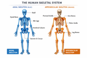
[Image: Labeled diagram of human skeletal system showing axial and appendicular skeleton
What Is the Skeletal System?
Definition (Exam-Oriented)
The skeletal system is the organ system composed of bones, cartilage, ligaments, and joints that provides structural support, protection to internal organs, facilitates movement, produces blood cells, and stores minerals.
An adult human skeleton consists of 206 bones, while newborns have around 270 bones, many of which fuse during growth.
Components of the Skeletal System
- Bones
- Cartilage
- Ligaments
- Joints
- Bone marrow
In an adult human, the skeletal system consists of 206 bones, though infants have more which later fuse.
Functions of the Skeletal System
From years of classroom experience, I always emphasize these core functions because exam questions frequently revolve around them:
- Support – Forms the structural framework of the body
- Protection – Safeguards vital organs (brain, heart, lungs, spinal cord)
- Movement – Acts as levers for muscles
- Mineral Storage – Reservoir for calcium and phosphorus
- Blood Cell Formation (Hematopoiesis) – Occurs in red bone marrow
- Fat Storage – Yellow bone marrow stores lipids
Divisions of the Skeletal System
From a teaching perspective, dividing the skeleton into two major parts helps students understand function through structure.
1. Axial Skeletal System
The axial skeleton forms the central axis of the body and is primarily concerned with support and protection.
It includes:
- Skull
- Vertebral column
- Ribs
- Sternum
- Number of bones: 80
2. Appendicular Skeletal System
The appendicular skeleton consists of bones of the limbs and girdles and is specialized for movement and locomotion.
It includes:
- Upper limbs
- Lower limbs
- Pectoral (shoulder) girdle
- Pelvic girdle
- Number of bones: 126
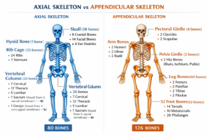
[Image: Side-by-side comparison diagram of axial and appendicular skeleton with labels and bone counts.
Also Read- General Principles of Cell Communication & Signaling
Types of Bones
Based on shape and structure, bones are classified into several types. This classification frequently appears in NEET, NCERT, and university exams.
1. Long Bones
Examples: Femur, humerus, tibia
Characteristics:
- Longer than wide
- Shaft (diaphysis) with expanded ends (epiphyses)
Functions:
- Act as levers for movement
- Support body weight
2. Short Bones
Examples: Carpals, tarsals
Characteristics:
- Cube-shaped
- Spongy bone inside
Functions:
- Provide stability with limited movement
3. Flat Bones
Examples: Skull bones, ribs, sternum, scapula
Characteristics:
- Thin and flattened
- Broad surface area
Functions:
- Protection of organs
- Muscle attachment
4. Irregular Bones
Examples: Vertebrae, facial bones
Characteristics:
- Complex shapes
Functions:
- Protection and support
5. Sesamoid Bones
Example: Patella
Characteristics:
- Embedded within tendons
Functions:
- Reduce friction
- Increase mechanical advantage
Salient Features and Functions of Axial Skeleton
1. Skull
The skull consists of 22 bones divided into:
- Cranial bones (8): Protect the brain
- Facial bones (14): Form the face
Functions:
- Protection of brain
- Formation of facial structure
- Support for sensory organs
2. Vertebral Column
Total vertebrae: 33 (26 in adults due to fusion)
Regions:
- Cervical (7)
- Thoracic (12)
- Lumbar (5)
- Sacral (5 fused)
- Coccygeal (4 fused)
Functions:
- Protects spinal cord
- Supports body weight
- Allows flexibility and posture
3. Thoracic Cage (Ribs & Sternum)
- Ribs: 12 pairs
- True ribs (1–7)
- False ribs (8–10)
- Floating ribs (11–12)
Functions:
- Protect heart and lungs
- Aid in respiration
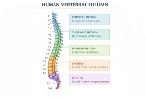
[Image: Detailed anatomical diagram of human vertebral column with cervical, thoracic, lumbar, sacral, and coccygeal regions labeled.
Also Read- Tissue Level of Organization: Structure and Functions
Salient Features and Functions of Appendicular Skeleton
1. Pectoral Girdle
Bones:
- Clavicle
- Scapula
Functions:
- Attach upper limbs to axial skeleton
- Provide wide range of motion
2. Upper Limbs
Bones include:
- Humerus
- Radius & Ulna
- Carpals, Metacarpals, Phalanges
Functions:
- Grasping and manipulation
3. Pelvic Girdle
Bones:
- Hip bones (ilium, ischium, pubis)
Functions:
- Attach lower limbs
- Protect pelvic organs
- Bear body weight
4. Lower Limbs
Bones include:
- Femur
- Tibia & Fibula
- Tarsals, Metatarsals, Phalanges
Functions:
- Locomotion
- Weight bearing
Organization of Skeletal Muscle
One mistake I often see in students is memorizing muscle terms without visualizing the hierarchy. Let’s fix that.
Structural Organization (From Large to Small)
- Muscle
- Fascicle
- Muscle fiber (muscle cell)
- Myofibril
- Sarcomere
- Myofilaments (actin & myosin)
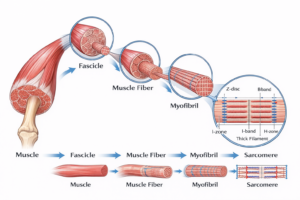
[Image: Step-by-step labeled diagram showing muscle → fascicle → muscle fiber → myofibril → sarcomere.
Connective Tissue Coverings
- Epimysium: Covers entire muscle
- Perimysium: Covers fascicles
- Endomysium: Covers muscle fibers
These layers provide strength, protection, and pathways for nerves and blood vessels.
Physiology of Muscle Contraction
Sliding Filament Theory
Muscle contraction occurs when actin filaments slide over myosin filaments, shortening the sarcomere without changing filament length.
Steps of Muscle Contraction
- Nerve impulse arrives
- Acetylcholine released
- Muscle action potential generated
- Calcium ions released from sarcoplasmic reticulum
- Actin-myosin cross-bridge formation
- Power stroke
- Muscle shortens
Role of ATP
ATP is required for:
- Cross-bridge formation
- Power stroke
- Detachment of myosin
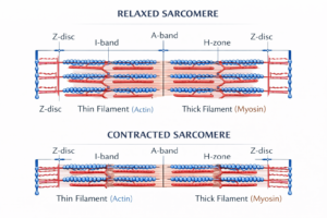
[Image: Educational diagram showing relaxed vs contracted sarcomere with actin and myosin interaction.
Neuromuscular Junction (NMJ)
Definition
The neuromuscular junction is the synapse between a motor neuron and a skeletal muscle fiber.
Structure of NMJ
- Presynaptic neuron terminal
- Synaptic cleft
- Motor end plate
Events at NMJ
- Nerve impulse reaches axon terminal
- Calcium enters neuron
- Acetylcholine released
- ACh binds to receptors
- Muscle action potential initiated
This is a high-yield exam topic, especially for physiology sections.
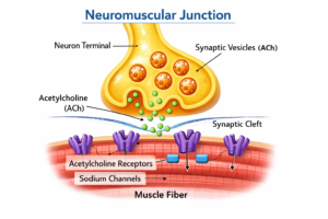
[Image: Clear labeled diagram of neuromuscular junction showing neuron terminal, synaptic cleft, acetylcholine receptors.
Clinical Correlation (Teaching Insight)
Conditions like:
- Myasthenia gravis (autoimmune NMJ disorder)
- Muscular dystrophy
- Osteoporosis
make much more sense when students truly understand skeletal and muscular physiology.
Unit 2 Human Anatomy And Physiology 1 – All Chapter PDF Notes
Frequently Asked Questions (FAQs)
Q1. How many bones are present in the adult human skeletal system?
Answer: 206 bones.
Q2. What is the main difference between axial and appendicular skeleton?
Answer: Axial skeleton provides support and protection, while appendicular skeleton enables movement.
Q3. Which type of bone is the femur?
Answer: Long bone.
Q4. What is the functional unit of a muscle fiber?
Answer: Sarcomere.
Q5. Which neurotransmitter is released at the neuromuscular junction?
Answer: Acetylcholine.
Conclusion: A Teacher’s Final Note
Having taught this topic for years, I can confidently say that the skeletal system becomes intuitive and fascinating when students see it as an integrated system rather than isolated facts. Bones, muscles, and nerves work together seamlessly to create movement, strength, and life itself.
Master these concepts deeply—not just for exams, but for understanding how your own body works every single day.