Structural and Functional Classification of Joints, Types of Joints Movements and Its Articulation
Introduction
When I teach joints to first-year medical, nursing, or pharmacy students, I always begin with one simple question:
“What is the point of having bones if they cannot move?”
That question naturally leads us to joints (articulations)—the most functionally significant connections in the human body. Understanding the structural and functional classification of Joints, types of joints movements and its articulation is not only essential for anatomy exams but also critical for clinical practice, physiotherapy, orthopedics, sports medicine, and rehabilitation.
In this article, I’ll explain joints the same way I’ve taught them over years in classrooms:
- Step-by-step
- With real anatomical examples
- Clinically relevant
- Exam-friendly
What Is a Joint (Articulation)?
A joint, also known as an articulation, is the site where two or more bones meet, or where a bone meets cartilage or teeth.
Primary Functions of Joints
- Provide movement
- Provide stability
- Transmit forces
- Protect underlying structures
Teaching Tip:
Students often memorize joint types but forget why joints exist. Always connect structure → movement → function.
Also Read- Structure and Functions of Cell | Transport, Division & Junctions
Structural Classification of Joints
Structural classification is based on:
- The material binding the bones
- The presence or absence of a joint cavity
There are three main structural types:
1. Fibrous Joints
In fibrous joints, bones are joined by dense connective tissue. There is no joint cavity, so movement is minimal or absent.
Types of Fibrous Joints
a) Sutures
- Found in the skull
- Bones are connected by short collagen fibers
- Example: Coronal suture, sagittal suture
Clinical Insight:
Sutures allow skull growth in infants. Premature fusion leads to craniosynostosis.
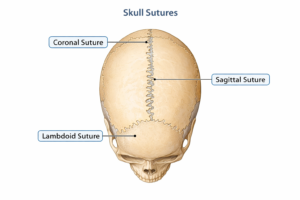
[Image: Labeled diagram of skull sutures – coronal, sagittal, lambdoid]
b) Syndesmosis
- Bones connected by ligaments or interosseous membrane
- Slight movement allowed
- Example: Tibia–fibula joint, radius–ulna
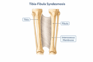
[Image: Tibia-fibula syndesmosis with interosseous membrane]
c) Gomphosis
- Peg-and-socket joint
- Example: Tooth in alveolar socket
2. Cartilaginous Joints
Bones are united by cartilage, allowing limited movement.
Types of Cartilaginous Joints
a) Primary Cartilaginous Joint (Synchondrosis)
- Cartilage type: Hyaline cartilage
- Temporary joints
- Example: Epiphyseal plate
Clinical Relevance:
Growth disorders directly affect these joints.
b) Secondary Cartilaginous Joint (Symphysis)
- Cartilage type: Fibrocartilage
- Permanent joints
- Example: Pubic symphysis, intervertebral discs
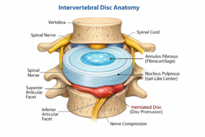
[Image: Intervertebral disc anatomy showing fibrocartilage]
3. Synovial Joints
These are the most complex and most mobile joints in the body.
Key Features of Synovial Joints
- Joint cavity
- Articular cartilage
- Synovial membrane
- Synovial fluid
- Fibrous capsule
- Ligaments
Teaching Insight:
If a joint moves freely, it is almost always synovial.
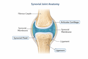
[Image: Labeled synovial joint showing capsule, synovial fluid, cartilage]
Also Read- General Principles of Cell Communication & Signaling
Functional Classification of Joints
Functional classification focuses on degree of movement, not structure.
1. Synarthrosis
- Immovable joints
- Example: Skull sutures
2. Amphiarthrosis
- Slightly movable joints
- Example: Pubic symphysis
3. Diarthrosis
- Freely movable joints
- All synovial joints
Exam Tip:
Diarthrosis = synovial joint = free movement.
Also Read- Tissue Level of Organization: Structure and Functions
Types of Synovial Joints (Based on Shape & Movement)
This is where students often struggle—but clarity comes from linking shape to movement.
1. Plane Joint
- Flat surfaces
- Gliding movement
- Example: Intercarpal joints
2. Hinge Joint
- Uniaxial movement
- Flexion and extension
- Example: Elbow, knee
3. Pivot Joint
- Rotation around a central axis
- Example: Atlas–axis joint
4. Condyloid (Ellipsoid) Joint
- Biaxial movement
- Example: Wrist joint
5. Saddle Joint
- Each surface is concave and convex
- Example: First carpometacarpal joint (thumb)
Clinical Insight:
Loss of saddle joint function severely affects grip.
6. Ball and Socket Joint
- Multiaxial movement
- Example: Shoulder, hip
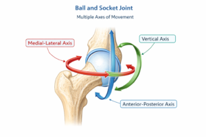
[Image: Ball and socket joint showing multiple axes of movement]
Also Read- Integumentary System: Structure and Functions of Skin
Types of Joint Movements
Understanding movements is essential for physiology, orthopedics, and physiotherapy.
Angular Movements
- Flexion
- Extension
- Abduction
- Adduction
Rotational Movements
- Medial rotation
- Lateral rotation
Special Movements
- Circumduction
- Supination & pronation
- Dorsiflexion & plantarflexion
- Inversion & eversion
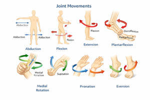
[Image: Diagram showing joint movements with arrows]
Also Read- Skeletal System: Bones, Muscles & Contraction Explained
Articulation: Functional Anatomy Perspective
Articulation is not just bone-to-bone contact. It involves:
- Muscles
- Ligaments
- Tendons
- Neural control
Clinical Relevance:
Joint disorders like arthritis affect articulation long before bones show damage.
Clinical Importance of Joint Classification
From years of teaching and clinical exposure, I can confidently say:
Structural classification helps diagnosis
Functional classification helps treatment planning
Movement analysis helps rehabilitation
Examples:
- Osteoarthritis → synovial joints
- Disc prolapse → cartilaginous joints
- Fracture dislocation → articulation failure
Unit 2 Human Anatomy And Physiology 1 – All Chapter PDF Notes
FAQ Section
What is meant by structural and functional classification of joints?
Structural classification is based on joint anatomy, while functional classification depends on movement allowed.
What are the main types of joint movements?
Joint movements include flexion, extension, abduction, adduction, rotation, and special movements.
Why are synovial joints freely movable?
Because they have a joint cavity filled with synovial fluid that reduces friction.
What is articulation in anatomy?
Articulation refers to the point where two bones meet and interact to allow movement.
Which joint allows the widest range of movement?
The shoulder joint (ball and socket joint).
Conclusion
A strong understanding of structural and functional classification of Joints, types of joints movements and its articulation forms the foundation of human movement science. Whether you are a student, educator, or healthcare professional, mastering these concepts will enhance your anatomical reasoning and clinical decision-making.